Contents

Source: Physik Instrumente
Fluorescence Microscopy: A Powerful Imaging Technique
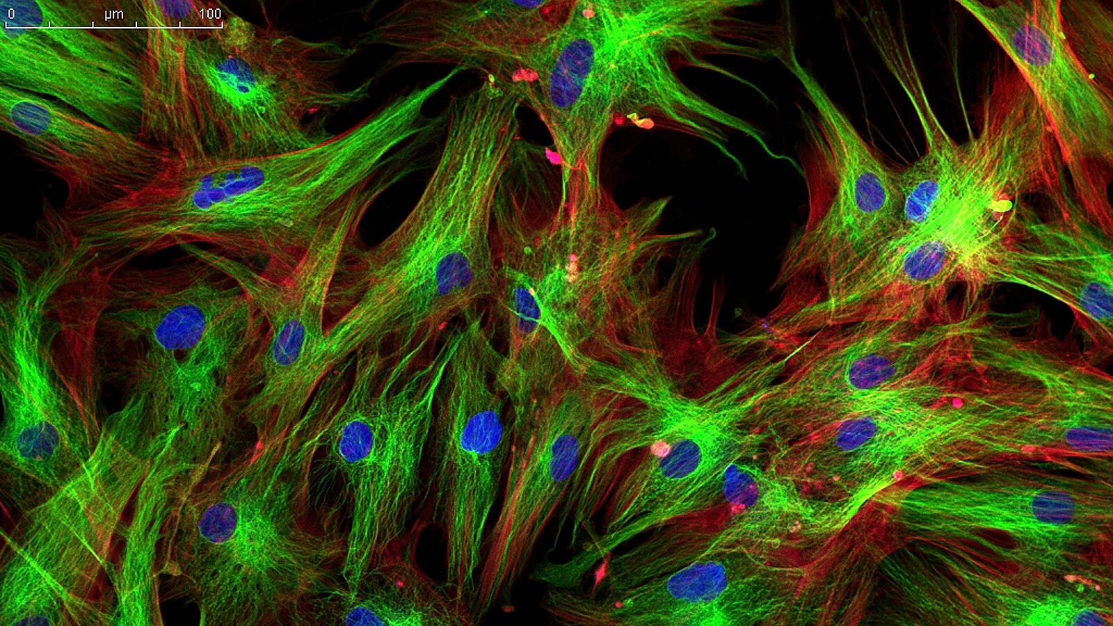
Fluorescence microscopy is a widely used technique for capturing microscopic images of samples, especially biological materials. It involves exciting the sample with a focused laser beam, scanning through the sample, collecting the fluorescent light emitted, and processing the data to generate images.
Confocal Geometry in Fluorescence Microscopy
In confocal fluorescence microscopy, the light from the focus in the sample is imaged onto a pinhole to enhance resolution and depth. This method allows for imaging relatively thick samples with high depth resolution, making it valuable in biomedical imaging.
Multiphoton Microscopy
Multiphoton microscopy is an advanced technique that uses femtosecond laser pulses for excitation. It offers improved resolution in both lateral and axial directions, better penetration of thicker samples, and reduced photobleaching effects compared to single-photon microscopy.
Stimulated Emission Depletion Microscopy
STED microscopy is a super-resolution technique that utilizes stimulated emission to achieve resolutions below the diffraction limit. This method, developed by Nobel laureates, allows imaging at the nanoscale level, surpassing traditional optical microscopy limits.
Stochastic Optical Reconstruction Microscopy
STORM is another super-resolution microscopy method that enables imaging below the diffraction limit by localizing individual fluorescent molecules. By combining multiple images, a complete high-resolution image of the sample can be reconstructed.
Applications of Fluorescence Microscopy
Fluorescence microscopy finds extensive applications in biological research, medical diagnosis, and studying living cells. It is particularly valuable in observing cellular processes without significant cell damage, making it a versatile tool in various scientific fields.
Conclusion
Fluorescence microscopy, with its various techniques like confocal, multiphoton, STED, and STORM, has revolutionized the field of microscopy by enabling high-resolution imaging beyond the diffraction limit. These methods have opened up new possibilities in biological research, nanoscale imaging, and medical diagnostics, pushing the boundaries of what was previously thought possible with optical microscopy.

Source: Bitesize Bio
Feel free to comment your thoughts.


